WITec Paper Award 2023 Recognizes Outstanding Publications
The 2023 Paper Award Winners
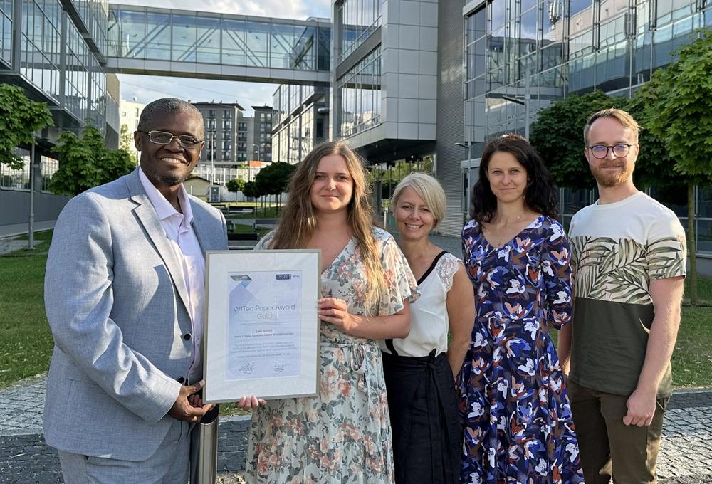
- GOLD: Ewa Stanek, Marta Z. Pacia, Agnieszka Kaczor, Krzysztof Czamara (2022) The distinct phenotype of primary adipocytes and adipocytes derived from stem cells of white adipose tissue as assessed by Raman and fluorescence imaging. Cellular and Molecular Life Sciences 79: 383. DOI: 10.1007/s00018-022-04391-2
- SILVER: Aïcha Badou, Sylvain Pont, Stéphanie Auzoux-Bordenave, Morgane Lebreton, Jean-François Bardeau (2022) New insight on spatial localization and microstructures of calcite-aragonite interfaces in adult shell of Haliotis tuberculata: Investigations of wild and farmed abalones by FTIR and Raman mapping. Journal of Structural Biology 214: 107854. DOI: 10.1016/j.jsb.2022.107854
- BRONZE: Stefano L. Oscurato, Francesco Reda, Marcella Salvatore, Fabio Borbone, Pasqualino Maddalena, Antonio Ambrosio (2022) Shapeshifting diffractive optical devices. Laser & Photonics Reviews 16: 2100514. DOI: 10.1002/lpor.202100514
For a list of all previous award-winning publications, please visit the official WITec Paper Award website.
The Paper Award GOLD: Characterization of cell development
As the world-wide incidence of obesity-related health issues increases, adipose tissue is of growing interest to researchers. In vitro investigations of biochemical processes require model cell lines that resemble the in vivo state as closely as possible and different protocols exist for obtaining model adipocytes from stem cells. Ewa Stanek and her colleagues Marta Z. Pacia, Agnieszka Kaczor and Krzysztof Czamara from the Jagiellonian Centre for Experimental Therapeutics and the Faculty of Chemistry of the Jagiellonian University (Kraków, Poland) win the Gold Paper Award 2023 for comparing primary and stem cell-derived adipocytes. The authors used Raman and fluorescence imaging for characterizing the maturation of adipocytes from brown and white adipose tissue. 2D and 3D Raman microscopy visualized changes in cell morphology and the distribution of lipids, DNA, and proteins that occur during adipogenesis. The formation of lipid droplets was observed and Raman spectral analysis enabled the quantification of lipid unsaturation levels. Comparing the results revealed no difference between stem cell-derived adipocytes from different tissue types. However, differences in chemical composition were found for primary adipocytes and those derived from stem cells. “Our study reveals limitations of existing model systems and that the chosen protocols can impact the experimental results,” observed Ewa Stanek. “These findings are relevant for metabolic studies and applications of tissue engineering.”
The Paper Award SILVER: Biomineralization of abalone shells
New materials for technological applications are often inspired by nature and researchers thus strive to understand how biomaterials are produced. The Silver Paper Award 2023 honors a study that investigates the biomineralization process of abalone shells, which was published by Aïcha Badou, PhD Engineer from the Muséum National d’Histoire Naturelle (MNHN, Station marine de Concarneau-DGD-REVE), and her colleagues Sylvain Pont, Stéphanie Auzoux-Bordenave, Morgane Lebreton and Jean-François Bardeau from the MNHN and Le Mans Université in France. The authors investigated the spatial distribution of the calcium carbonate (CaCO3) polymorphs aragonite and calcite in abalone shells with Raman imaging and Fourier-transform infrared spectroscopy (FTIR). Detailed analysis of the Raman spectra distinguished zones of differing degrees of order within areas of the same polymorph. Additionally, scanning electron microscopy (SEM) analyzed the shells’ microstructure and energy-dispersive X-ray spectroscopy (EDS) detected trace elements in the different zones. Based on their results, the authors proposed a model for microstructure and layer composition in relation to the direction of shell growth in thickness and length. “Our model explains different observations regarding shell structure reported in the past. The findings highlight the need for accurate sample documentation in order to ensure comparability,” said Dr. Badou. Future studies will investigate the influence of environmental factors on the growth patterns.
The Paper Award BRONZE: Development of shapeshifting planar optical elements
From microbiology to astronomy, imaging systems are important tools in various fields of research. Depending on their intended application, they contain different optical elements for structuring and manipulating light. Stefano L. Oscurato from the University of Naples, Italy, wins the Bronze Paper Award 2023 for his work on shapeshifting optical devices together with his colleagues Francesco Reda, Marcella Salvatore, Fabio Borbone, Pasqualino Maddalena and Antonio Ambrosio. The authors created planar diffractive optical elements, such as lenses and gratings, using a photoresponsive polymer that can be shaped into a desired morphology by incident light. The morphology defines the devices’ optical properties and was verified by atomic force and scanning electron microscopy. A custom-built holographic setup was able to shape the polymer surface, and visualize the resulting diffraction properties in real-time. The same polymer surface can be reshaped repeatedly while remaining in the beam path. “There are many applications that can profit from the ability to change imaging parameters without physically exchanging or moving optical elements,” commented Dr. Oscurato. Examples described in the paper include: gratings with variable diffraction properties, and a diffractive lens with an adjustable focal length that could serve to alter the magnification of a microscope.
The WITec Paper Award 2024 is open for submissions
The WITec jury looks forward to receiving many exceptional submissions for the WITec Paper Award 2024. Articles are eligible if they were published in 2023 (in print or online) in a peer-reviewed journal and contain results obtained with a WITec instrument. Send your publications as a PDF to papers@WITec.de before January 31, 2024, for a chance to win.
WITec GmbH pioneered 3D Raman imaging and correlative microscopy and continues to lead the industry with a product portfolio that offers speed, sensitivity and resolution without compromise. Raman, AFM and SNOM microscopes, select combinations thereof, and WITec-developed Raman-SEM (RISE) instruments can be configured for specific challenges in chemical and structural characterization through a modular hardware and software architecture with built-in capacity for expansion. Research, development and production are located at WITec headquarters in Ulm, Germany, and the WITec sales and support network has an established presence in every global region. In September 2021, WITec became a member of the Oxford Instruments Group, bringing technology leadership in Raman microscopy to its extensive portfolio of businesses.
WITec GmbH
Lise-Meitner-Str. 6
89081 Ulm
Telefon: +49 (731) 14070-0
Telefax: +49 (731) 14070-200
https://Raman.oxinst.com
Technical Marketing & PR
Telefon: +49 731 140 700
Fax: +49 173 140 70 200
E-Mail: eleni.vomhof@witec.de
![]()